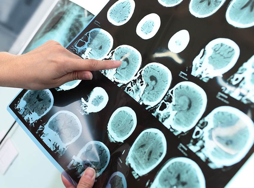
The human brain has three major components. The main part is the cerebrum surrounded by an outer layer known as the cerebral cortex. This part of the brain is required for remembering, problem solving, feeling and thinking. At the back of the brain is the cerebellum, important for coordination and balance. The brain stem is the connection between the brain and spinal cord and controls automatic functions, such as breathing, heartbeat and blood pressure.
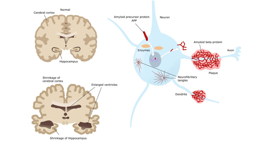
Our brains have an extensive blood vessel network to supply the more than 1000 billion neurons (nerve cells) that form the ‘neuron forest’ within our brain. Each of these neurons has multiple extended branches that connect and communicate with other neurons via small electrical pulses known as synapses, resulting in the release of tiny neurotransmitter molecules. In the brains of Alzheimer’s disease patients, these neurotransmitters, synapses and neurons are gradually destroyed and the brain slowly shrinks as the cells die. The hippocampus is usually the initial brain region affected in Alzheimer’s patients. The hippocampus is important for the formation of new memories; hence the reason that the first sign of Alzheimer’s is usually memory loss. When an affected brain is observed under the microscope, the brain shrinkage and decreased neurons and synapses are easily observed, along with protein accumulations within and between the neurons. It is still unclear whether these protein accumulations are actually the cause of Alzheimer’s disease or whether they are a by-product of other changes within the brain. However, it is known that these protein accumulations interfere with the synapses between neurons and prevent the release of essential neurotransmitters, resulting in characteristic cell death and brain shrinkage.
Amyloid Plaques The protein accumulations observed between the cells in the brain of Alzheimer’s disease patients are known as amyloid plaques and are formed from abnormal deposits of the beta-amyloid protein. The amyloid precursor protein is a normal protein present in the cell membrane of healthy neurons. When the amyloid precursor protein is cleaved by the alpha-secretase enzyme, the resulting protein is beneficial to neuron growth and survival. However, if it is instead cleaved by beta-secretase and gamma-secretase, the resulting protein is beta-amyloid. As the concentration of beta-amyloid increases, it forms into oligomers composed of 2-12 copies of the protein. The oligomers clump together and grow larger, incorporating other proteins to form the characteristic pathogenic plaques found between the neurons of individuals with Alzheimer’s disease. These plaques stop the synapses between the neurons and may also activate a chronic immune response, leading to a damaging inflammatory response.
Tau Tangles The protein accumulations within the cells in the brain of individuals affected by Alzheimer’s disease are known as tau tangles (or neurofibrillary tangles). The normal tau protein helps support the essential microtubule transport system within the cells. This transport system is like a set of train tracks and is essential to move nutrients, molecules and cell components within the cell. When abnormal tau is formed, it fails to maintain this transport system and the microtubules collapse, twist and disintegrate. The cell is unable to move molecules as required and eventually shrinks and dies.
Other Changes There are several other pathogenic changes that occur in the brain of an Alzheimer’s patient. These include an impaired glucose metabolism and carbohydrate regulation associated with insulin resistance, oxidative stress, inflammation, and defective copper regulation. These pathogenic changes are generally worse in the regions of the brain that show the most shrinkage and damage when viewed under the microscope.
References:
- Alzheimer’s Disease Fact Sheet. (Page Last Updated: July 20, 2015).
- Alzheimer’s Disease Education and Referral (ADEAR) Center – A service of the National Institute on Aging, National Institutes of Health.Bird TD (1998).
- Alzheimer Disease Overview.[Updated 2015 Sep 24]. In: Pagon RA, Adam MP, Ardinger HH, et al., editors. GeneReviews® [Internet]. Seattle (WA): University of Washington, Seattle; 1993-2015. Xu I et al. (2016).
- Elevation of brain glucose and polyol-pathway intermediates with accompanying brain-copper deficiency in patients with Alzheimer’s disease: metabolic basis for dementia. Scientific Reports 6: 27524.

